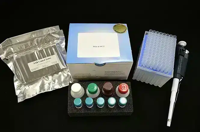
Microcystin BX Test Kit (ELISA Plate)
INTENDED USE
The Microcystin BX Plate Kit is a competitive ELISA for the quantitation of Microcystins in water.
REAGENTS AND MATERIALS PROVIDED
MATERIALS PROVIDED
• Plate – (1) containing 12 strips of 8 wells coated with sheep anti-rabbit antibodies
• Negative Control – (1) vial containing 2 mL of 0.0 ppb (µg/L) Microcystin-LR
• Microcystin BX Calibrators – (4) vials containing 2mL with a concentration of 0.1, 0.3, 0.8 and 2.0 ppb of Microcystin-LR
• Positive Control – (1) vial containing 2 mL of 1.0 ppb Microcystin-LR control
• Microcystin BX HRP Enzyme Conjugate – (1) bottle containing 8 mL
• Anti-Microcystin BX Antibody Solution – (1) bottle containing 8 mL
• Substrate – (1) bottle containing 14 mL
• Stop Solution – (1) bottle containing 14 mL
• 100X Wash Solution – (1) bottle containing 25 mL (Must be diluted before use. See Assay Procedure Step 3.)
SKU:
BAS-20-0300
Categories: Algal Toxins, Environmental Testing, Water
Description
USE PRINCIPLES
The Microcystin BX Plate Kit uses a polyclonal antibody that binds both Microcystins and a Microcystin-enzyme conjugate. Microcystins in the sample compete with the Microcystin enzyme conjugate for a limited number of antibody binding sites. In the assay procedure you will:
• Add Microcystin-enzyme conjugate and calibrator or sample containing Microcystins to a test well, followed by antibody solution. The conjugate competes with any Microcystins in the sample for the same antibody binding sites. The test well is coated with anti-rabbit IgG to capture the rabbit anti-Microcystin added.
• Wash away any unbound molecules, after you incubate this mixture for 30 minutes.
• Add colorless substrate solution to each well. In the presence of bound Microcystin-enzyme conjugate, the substrate is converted to a blue compound. One enzyme molecule can convert many substrate molecules.
Since the same number of antibody binding sites are available in every well, and each well receives the same number of Microcystin-enzyme conjugate molecules, a sample containing a low concentration of Microcystins allows the antibody to bind to many Microcystin-enzyme conjugate molecules. The result is a dark blue solution. Conversely, a high concentration of Microcystins allows fewer Microcystin-enzyme conjugate molecules to be bound by the antibodies, resulting in a lighter blue solution.
NOTE: Color is inversely proportional to Microcystin BX concentration.
Darker color = Lower concentration
Lighter color = Higher concentration
PRECAUTIONS
- Store all kit components at 4°C to 8°C (39°F to 46°F) when not in use.
- Do not freeze kit components or expose them to temperatures greater than 37°C (99°F).
- Allow all reagents and samples to reach ambient temperature before you begin the test.
- Do not use kit components after the expiration date.
- Do not mix reagents or test strips from kits with different lot numbers.
- Transfer of samples and reagents by pipette requires constant monitoring of technique. Pipetting errors are the major source of error in immunoassay methodology.
Additional information
| Format: |
96-well microtiter plate (12 test strips of 8 wells) |
|---|---|
| Standards: |
0 ,0.10 ,0.30 ,0.80 ,2 ppb |
| Incubation Time: |
60 Minutes |
Shipping & Delivery


