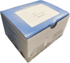
Microcystin Test Kit (Tube)
INTENDED USE
The Microcystin Tube Kit is an immunological laboratory test for the quantitation of Microcystins in water.
REAGENTS AND MATERIALS PROVIDED
MATERIALS PROVIDED
2 Bags Antibody coated tubes – each containing 20 tubes
1 vial of Negative Control containing 5 ml of 0.0 ppb Microcystin-LR
1 vial of Microcystin Calibrators – containing 5ml with a concentration of 0.3, 0.8, 2.0 and 5.0 ppb of Microcystin-LR
1 vial of Positive Control – containing 5 mL of 1.0 ppb Microcystin-LR control
1 bottle of Microcystin-HRP Enzyme Conjugate, containing 24 ml
1 bottle of Microcystin Antibody Solution – containing 24 ml
1 bottle of Substrate – containing 25 ml
1 bottle of Stop Solution – containing 25 ml
1 bottle of 100X Wash Solution – containing 25 ml (Must be diluted before use, See Assay Procedure Step 3)
SKU:
BAS-20-0098
Category: Algal Toxins
Description
SPECIFICITY
The Microcystin Tube Kit can detect several microcystin congeners. The % cross reactivity (% CR) of microcystin, other microcystin congeners relative to microcystin-LR is shown in the table below.
Congeners % CR
Microcystin-LR 100%
Microcystin-RR 73%
Microcystin-YR 58%
Microcystin-LA 2%
Microcystin-LF 3%
Microcystin-LW 4%
Nodularin 126%
USE PRINCIPLES
The Microcystin Tube Kit uses a polyclonal antibody that binds both Microcystins and a Microcystin-enzyme conjugate. Microcystins in the sample compete with the Microcystin enzyme conjugate for a limited number of antibody binding sites. In the assay procedure you will:
• Add Microcystin-enzyme conjugate and a sample containing Microcystins to a test well, followed by antibody solution. The conjugate competes with any Microcystins in the sample for the same antibody binding sites. The test tube is coated with anti-rabbit IgG to capture the rabbit anti-Microcystin added.
• Wash away any unbound molecules, after you incubate this mixture for 20 minutes.
• Add colorless substrate solution to each well. In the presence of bound Microcystin-enzyme conjugate, the substrate is converted to a blue compound. One enzyme molecule can convert many substrate molecules.
Since the same number of antibody binding sites are available in every tube, and each tube receives the same number of Microcystin-enzyme conjugate molecules, a sample containing a low concentration of Microcystins allows the antibody to bind to many Microcystin-enzyme conjugate molecules. The result is a dark blue solution. Conversely, a high concentration of Microcystins allows fewer Microcystin-enzyme conjugate molecules to be bound by the antibodies, resulting in a lighter blue solution.
NOTE: Color is inversely proportional to Microcystin concentration.
Darker color = Lower concentration
Lighter color = Higher concentration
PRECAUTIONS
• Store all kit components at 4°C to 8°C (39°F to 46°F) when not in use.
• Each reagent is optimized for use in the Microcystin Tube Kit. Do not substitute reagents from any other manufacturer into the test kit. Do not combine reagents from other Microcystin Tube Kit with different lot numbers.
• Dilution or adulteration of reagents or samples not called for in the procedure may result in inaccurate results.
• Do not use reagents after expiration date.
• Reagents should be brought to room temperature, 20 to 28ºC (62 to 82ºF) prior to use. Avoid prolonged (> 24 hours) storage at room temperature.
• The Stop Solution is 1N hydrochloric acid. Avoid contact with skin and mucous membranes. Immediately clean up any spills and wash area with copious amounts of water. If contact should occur, immediately flush with copious amounts of water.
• Transfer of samples and reagents by pipette requires constant monitoring of technique. Pipetting errors are the major source of error in immunoassay methodology.
• Use approved methodologies to confirm and positive results.
Additional information
| Format: |
40 antibody coated test tubes |
|---|---|
| Standards: |
0 ,0.30 ,0.80 ,2 ,5 ppb |
| Incubation Time: |
40 Minutes |
Shipping & Delivery


